Application to non-invasive biomedical measurements such as cancer cell detection and treatment
Diagnostic Imaging of Breast Cancer Cells
X-ray imaging is the mainstay of breast cancer diagnosis in Japan and abroad. However, due to radiation exposure and breast compression, the consultation rate is only about 10-20%, even though it is the most common type of cancer, and the false positive rate (the probability of a positive diagnosis in the absence of cancer) is reported to be more than 50% because it is difficult to identify early cancers smaller than 10 mm. The false-positive rate (the probability of a positive diagnosis in the absence of cancer) is reported to be as high as 50% or more.
On the other hand, microwave breast cancer imaging is promising as a simpler and more frequent breast cancer screening technique because it uses electromagnetic waves in the safe wavelength band used in cell phones and wifi, and the equipment is compact and inexpensive. In particular, it has been confirmed that the complex permittivity of cancer tissue is more than ten times higher than that of fat and other tissues, and that three-dimensional imaging of the complex permittivity distribution can be used to detect early cancers of less than 10 mm in size, which is difficult to detect using X-rays or ultrasound.
In microwave cancer diagnosis, either the radar method, in which imaging is performed from the reflected signal from the target, or the tomography method, in which the complex permittivity is directly reconstructed from the scattered signal, is employed, taking advantage of the fact that the conductivity of cancer cells is significantly different from that of normal cells.
In our laboratory, we focus on the tomography method, which can directly reconstruct the complex permittivity. The tomography method requires a large amount of computation time for sequential problem analysis, etc. The CSI (Contrast Source Inversion) method is a method that avoids this problem. This method solves the data equation and two integral equations called the state equation simultaneously, and it is known that it can ensure accuracy even for nonlinear problems.
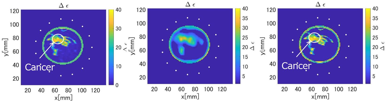
On the other hand, to maintain sufficient resolution, the image pixel size must be small. In this case, a large number of unknowns must be set, which is larger than the number of observed data, and thus requires solving a bad set problem whose solution cannot be uniquely determined, which limits the resolution. Focusing on the reconstruction of only mammary gland and cancer tissues with high dielectric constant, we have devised an algorithm that does not assign unknowns to fat and other tissues, and have proposed a method to improve resolution by greatly reducing the number of unknowns. Figure 1 shows the results of reconstruction using conventional CSI and ROI-restricted CSI. The results show that the proposed method provides higher resolution and estimates the dielectric constant of cancer cells with higher accuracy than conventional methods, and is significantly more useful for cancer cell identification.
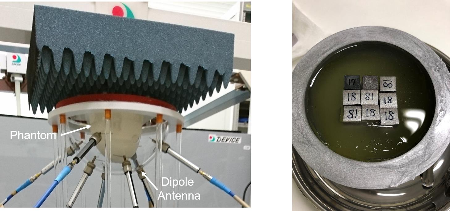
Currently, we are validating the effectiveness of these methods using phantoms and clinical data from a microwave mammography system as shown in Figure 2.
Cancer Treatment Application Using Microwave Ablation (Cauterization)
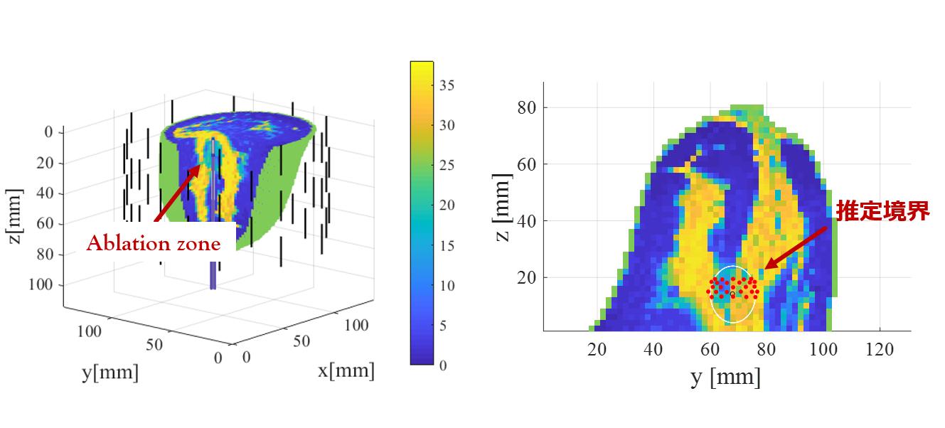
Biological tissue absorbs microwave energy as heat due to its high electrical conductivity. Therefore, irradiating microwaves to the area where cancer cells exist using a probe can cauterize the area and kill the cancer cells. This treatment method, called microwave ablation (MWA), is attracting attention as a promising cancer treatment method because it can reliably remove cancer cells without surgery. On the other hand, the energy level must be adjusted by sequentially monitoring the area to avoid ablation of normal cells other than cancer cells. In general, the dielectric constant is temperature-dependent, and it is reported that the dielectric constant and conductivity decrease to 60 to 70% of their original values when the tissue temperature reaches close to 100 degrees. In our laboratory, the dielectric constant of the ablation region is measured by the ablation method. We have proposed a method to measure the ablation region in real time using an effective signal processing method.
Figure 3 shows the result of the monitoring developed in our laboratory, in which the ablation area is estimated with high accuracy. This processing time can be achieved within 0.1 second on an ordinary PC, and we believe it will contribute greatly to the realization of this treatment technology.
Kazuki Noritake and Shouhei Kidera,
"
Boundary Extraction Enhanced Distorted Born Iterative Method for Microwave Mammography
",
IEEE Antennas and Wireless Propagation Letters., Vol.18, NO.4, pp.776-780, Apr., 2019.
H. Sato and S. Kidera,
" Multi-frequency Integration Algorithm of Contrast Source Inversion Method for Microwave Breast Tumor Detection"
41st IEEE International Engineering in Medicine and Biology Conference 2019 (IEEE EMBC 2019), Berlin, Germany, July, 2019. K. Kanazawa and S. Kidera,
" Experimental Validation for Microwave Based Real-time Monitoring for Microwave Ablation Treatment"
41st IEEE International Engineering in Medicine and Biology Conference 2019 (IEEE EMBC 2019), Berlin, Germany, July, 2019.
X-ray imaging is the mainstay of breast cancer diagnosis in Japan and abroad. However, due to radiation exposure and breast compression, the consultation rate is only about 10-20%, even though it is the most common type of cancer, and the false positive rate (the probability of a positive diagnosis in the absence of cancer) is reported to be more than 50% because it is difficult to identify early cancers smaller than 10 mm. The false-positive rate (the probability of a positive diagnosis in the absence of cancer) is reported to be as high as 50% or more.
On the other hand, microwave breast cancer imaging is promising as a simpler and more frequent breast cancer screening technique because it uses electromagnetic waves in the safe wavelength band used in cell phones and wifi, and the equipment is compact and inexpensive. In particular, it has been confirmed that the complex permittivity of cancer tissue is more than ten times higher than that of fat and other tissues, and that three-dimensional imaging of the complex permittivity distribution can be used to detect early cancers of less than 10 mm in size, which is difficult to detect using X-rays or ultrasound.
In microwave cancer diagnosis, either the radar method, in which imaging is performed from the reflected signal from the target, or the tomography method, in which the complex permittivity is directly reconstructed from the scattered signal, is employed, taking advantage of the fact that the conductivity of cancer cells is significantly different from that of normal cells.
In our laboratory, we focus on the tomography method, which can directly reconstruct the complex permittivity. The tomography method requires a large amount of computation time for sequential problem analysis, etc. The CSI (Contrast Source Inversion) method is a method that avoids this problem. This method solves the data equation and two integral equations called the state equation simultaneously, and it is known that it can ensure accuracy even for nonlinear problems.
On the other hand, to maintain sufficient resolution, the image pixel size must be small. In this case, a large number of unknowns must be set, which is larger than the number of observed data, and thus requires solving a bad set problem whose solution cannot be uniquely determined, which limits the resolution. Focusing on the reconstruction of only mammary gland and cancer tissues with high dielectric constant, we have devised an algorithm that does not assign unknowns to fat and other tissues, and have proposed a method to improve resolution by greatly reducing the number of unknowns. Figure 1 shows the results of reconstruction using conventional CSI and ROI-restricted CSI. The results show that the proposed method provides higher resolution and estimates the dielectric constant of cancer cells with higher accuracy than conventional methods, and is significantly more useful for cancer cell identification.
Currently, we are validating the effectiveness of these methods using phantoms and clinical data from a microwave mammography system as shown in Figure 2.
Figure 1: Results of breast phantom reconstruction using the CSI method (left: true values, center: conventional CSI, right: CSI with ROI restriction)
Figure 2: Microwave mammography system (left) and breast phantom (right)
Cancer Treatment Application Using Microwave Ablation (Cauterization)
Biological tissue absorbs microwave energy as heat due to its high electrical conductivity. Therefore, irradiating microwaves to the area where cancer cells exist using a probe can cauterize the area and kill the cancer cells. This treatment method, called microwave ablation (MWA), is attracting attention as a promising cancer treatment method because it can reliably remove cancer cells without surgery. On the other hand, the energy level must be adjusted by sequentially monitoring the area to avoid ablation of normal cells other than cancer cells. In general, the dielectric constant is temperature-dependent, and it is reported that the dielectric constant and conductivity decrease to 60 to 70% of their original values when the tissue temperature reaches close to 100 degrees. In our laboratory, the dielectric constant of the ablation region is measured by the ablation method. We have proposed a method to measure the ablation region in real time using an effective signal processing method.
Figure 3 shows the result of the monitoring developed in our laboratory, in which the ablation area is estimated with high accuracy. This processing time can be achieved within 0.1 second on an ordinary PC, and we believe it will contribute greatly to the realization of this treatment technology.
Figure 3: Example of microwave ablation area estimation
References
" Multi-frequency Integration Algorithm of Contrast Source Inversion Method for Microwave Breast Tumor Detection"
41st IEEE International Engineering in Medicine and Biology Conference 2019 (IEEE EMBC 2019), Berlin, Germany, July, 2019.
" Experimental Validation for Microwave Based Real-time Monitoring for Microwave Ablation Treatment"
41st IEEE International Engineering in Medicine and Biology Conference 2019 (IEEE EMBC 2019), Berlin, Germany, July, 2019.